The Who, What, and When of an MRI
What is an MRI?
MRI stands for Magnetic Resonance Imaging. To explain how it works can be difficult, but I will try to break it down into its simplest form. The MRI uses magnetic fields and computer-generated radiofrequency waves to create detailed images of the organs and tissues in your body. The magnetic field temporarily realigns water molecules in your body. Radiofrequency waves cause these aligned atoms to produce faint signals, which are used to create cross-sectional MRI images — like slices in a loaf of bread. The “slice” can occur in three different planes ( i.e., front to back- frontal, side to side – sagittal, cross-sectional – transverse)
What can an MRI show the vet, and why is it helpful?
An MRI in a horse offers a couple of significant advantages over other forms of imaging. First of all, an MRI can evaluate both bone and soft tissue structures. Whereas radiology or X-rays are primarily for bone, an ultrasound is primarily for soft tissue, and nuclear medicine/bone scan is primarily for bone. Secondly, an MRI can image the structures of the foot extremely well, which is a common source of pain and lameness in the horse. We can generate X-rays for the bones of the foot adequately, but we cannot visualize the soft tissue structures of the foot very well because of the hoof wall, but the MRI allows us to do so. Thirdly, an MRI and CT are considered 3-dimensional imaging forms, while radiographs, ultrasounds, and nuclear medicine are only 2- 2-dimensional imaging. Three-dimensional imaging gives us a much greater detail of the location and status of normal and diseased tissues.
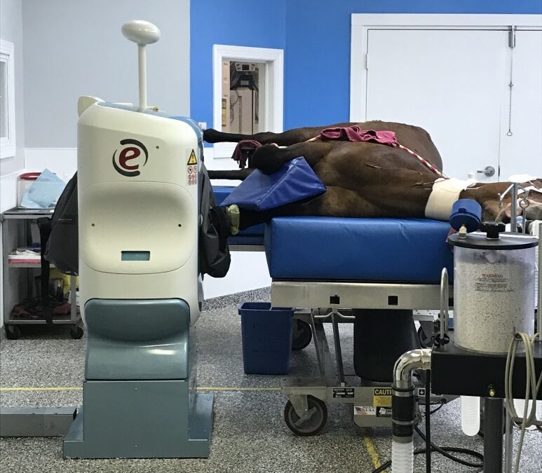
Who is a good candidate for an MRI?
While there are various reasons a horse may be referred for MRI, the most common reason is lameness. The lameness could range from a low-grade intermittent to persistent lameness or acute one. At BREC, we can perform MRI studies from the carpus and hock down to the foot. Our magnet (Esoate-O Scan) was originally designed to do human extremities (i.e., hands, feet, legs, and arms). We have a very small radiofrequency coil, which allows for a lot less signal “noise” and creates a better image quality. Unfortunately, because we have a small radiofrequency coil, we cannot perform studies on draft horses or horses with potentially very large feet.
When would an MRI be something an owner should consider doing?
There are many reasons for your horse to get an MRI, aside from the amount of time and money that can be lost from an inaccurate diagnosis and inappropriate treatments, and don’t forget about lost training time! One of the biggest reasons to get an MRI is it offers the most definitive diagnosis, which will then allow you and your veterinarian to formulate a comprehensive treatment and rehabilitation plan. Furthermore, a follow-up MRI can also be used to assess healing and response to therapy.
What should be done before an MRI can be scheduled?
Keep in mind that we cannot MRI a whole body or, for that matter, a whole leg. With the MRI, we have roughly a 5-6 inch “field of view” or study. So, it is crucial to have a lameness examination performed and nerve and/or joint blocks to localize the region of lameness. This will determine which fields of view should undergo an MRI. This also helps interpret the significance of the MRI findings.
By Dr. Steven S Trostle DVM, MS, DACVS, DACVSMR